Tuesday, January 3, 2012

Neuroimaging Primer


Neuroimaging Primer

Keith A. Johnson, M.D., Harvard Medical School

page 1 2
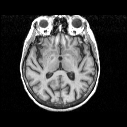
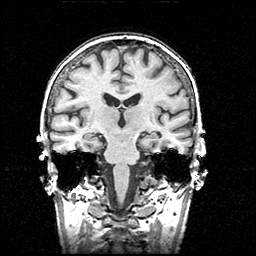
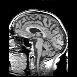
Tome is Greek for slice. The standard slice orientation in most brain imaging is transaxial or "axial". Left is shown at right. Note that, like the "lower organs", we look up to the brain. Other standard planes of view arecoronal and sagittal. Non-tomographic images represent "projections" from a single point of view and include bolus contrast x-ray angiograms and MR angiograms.
Tomographic images are made up of little squares called "pixels" (picture elements), each of which takes a grey-scale value from 1 (black) to 256 (white). Each pixel represents brain tissue which is about 1 mm. on each of two sides. The thickness of the slice is often 3 or 5 mm, thus creating a three-dimensional volume element, or "voxel", which is shaped like a shoe box. Pixel intensity represents an average from tissue within the voxel.

Image types

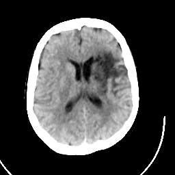
  • CT (roentgen-ray computed tomography) A beam of x-rays is shot straight through the brain. As it comes out the other side, the beam is blunted slightly because it has hit dense living tissues on the way through. Blunting or "attenuation" of the x-ray comes from the density of the tissue encountered along the way. Very dense tissue like bone blocks lots of x-rays; grey matter blocks some and fluid even less. X-ray detectors positioned around the circumference of the scanner collect attenuation readings from multiple angles. A computerized algorithm reconstructs an image of each slice. (example)
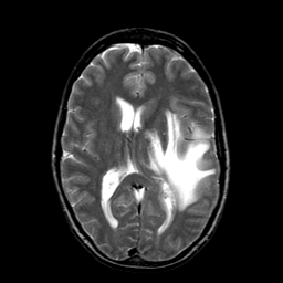
  • MRI (magnetic resonance imaging) When protons (here brain protons) are placed in a magnetic field, they become capable of receiving and then transmitting electromagnetic energy. The strength of the transmitted energy is proportional to the number of protons in the tissue. Signal strength is modified by properties of each proton's microenvironment, such as its mobility and the local homogeneity of the magnetic field. MR signal can be "weighted" to accentuate some properties and not others.When an additional magnetic field is superimposed, one which is carefully varied in strength at different points in space, each point in space has a unique radio frequency at which the signal is received and transmitted. This makes constructing an image possible. It represents the spatial encoding of frequency, just like a piano. (example). More details of MR here.
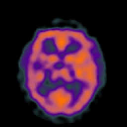
  • SPECT/PET (single photon/positron emission computed tomography) When radiolabeled compounds are injected in tracer amounts, their photon emissions can be detected much like x-rays in CT. The images made represent the accumulation of the labeled compound. The compound may reflect, for example, blood flow, oxygen or glucose metabolism, or dopamine transporter concentration. Often these images are shown with a color scale. (example)

MMed Anatomy, Spotter


Neuroanatomy is NB
Thoracic Outlet Syndrome

  • Thoracic inlet syndrome
  1. 1
    1. Azygos vein at level T4
      1. Lable diagram cross section T4 / T3 (muscles and bones) (20)
    2. Trachea
    3. Right Vein Pulmonary Trunk
      1. Pretracheal Lymphnode
  2. 1
    1. Hemiazygos vein
      1. Branch T9 communicates with Azygos system
    2. Internal Thoracic artery
    3. Vagus nerve left
      1. Compare to left phrenic nerve
  3. 1
    1. Left Main Bronchus as it enters the hilus
    2. Vertebral Artery left
    3. Subclavian Artery Right
  4. 1
    1. Right Descending Coronary Artery
      1. Compare anatomy of all structures of the heart - table
    2. Moderator band
      1.  Conduction in Right ventricle
    3. Infundibulum Right Pulmonary Artery
      1. Discuss essay - illustrate draw
  5. 1
    1. Scalenus Medius Mm - to the 2nd Rib
    2. Inferior Root of Brachial Plexus at T1
    3. Subclavian Artery Left
  6. 1
    1. Esophagus
    2. Caval Opening of the Inferior Vena Cava at T8
    3. Inferior Left Secondary Bronchus after Hilar Bifurcation
  7. 1
    1. Pectinate Muscle Rt
    2. Left Auricle
    3. Trabeculae carneae
  8. 1
    1. Middle Coronary vein 
    2. Ventricular Septum
    3. Coronary Sinus
  9. 1
    1. Phrenic Nerve
    2. Vagus Nerve - Recurrent laryngeal - Look at the Right Subclavian Artery with Recurrent Laryngeal Nerve
      1. The RIGHT RECURRENT LARYNGEAL NERVE loops beneath the arch of the subclavian artery to recurr (ascend) to the larynx of which it supplies. 
      2. Hoarseness
    3. Ligamentum Arteriosum (6)
      1. Dog heart 2.jpg
  10. 1
    1. ?? Brachiocephalic Artery
    2. Phrenic Nerve Left
      1. Look for Vagus nerve
    3. Left Coronary carotid Artery
  11. 1
    1. Brachiocephalic Vein left over aorta
    2. Left Vagus Nerve
    3. Nn Sympathetic Chain 
      1. Thoracic splanchnic nerves (greater, lesser, and least)
        1. File:Gray839.png
  12. 1
    1. Heart - Left Interventricular Cardiac Artery
    2. Papillary Muscle
    3. Chordae tendineae
      1.  
  13. 1
    1. Mm Innermost Thoracic Muscle Left
    2. Nn Intercostal Nerve T9  
    3. Nn Sympathetic Chain with branches to the Splantic Nerve
  14. 3x
    1. Left Phrenic Nerve C3,C4, C5 - referred pain to the left shoulder
    2. Azygos Vein
    3. Pulmonary Trunk
  15. 3x
    1. Vv Esophageal Veins
      1.  
    2. Inferior left main bronchus
    3. Intercostal 5th Artery from the Aorta
  16. 3x
    1. Falciform Ligament of the Liver
      1. File:Gray1085.png
    2. Pulmonary Vein
    3. Peritonium Abdomen
  17. 2x
    1. Left Scalenus anterior
      1. Scalenus anterior.png
      2. No chance in hell that we will see a subclavicular muscle - too difficult to dissect 
    2. Nn Vagus
      1. File:Gray793.png
  18. 2x
    1. Right central tendon of Diaphragm
    2. Caval opening at t8
      1. File:Gray391.png
  19. 3x
    1. Acromial end of left clavicle
    2. C2 odontoid process (dens) of cervical vertebra
      1. Gray87.png
    3. Thoracic vertebrae 
      1. There are no transverse foramen like the cervical vertebrae
      2. Gray82.png
  20. 1
    1. Rib typical - 2 articular facets
    2. Sternal notch
    3. 1st Rib left
  21. 1
    1. Xiphoid Process - T10
    2. Posterior arch of Atlas
    3. C3, C4, C5, C6 have transverse process
  22. 1
    1. C7 Transverse process
    2. T1
    3. Thoracic vertebrae
    4. Lumber vertebra
  23. 1
    1. Arteriogram
      1. Left Common Carotid Artery
      2. Dorsal Scapula Artery and Subcostal Artery
      3. Suprascapular Artery
      4. Anastomosis
    2. T2 Cross-section
    3. T4, T5, T10
    4. T4 Azygos vein

    11


    2 February 2011, Spot Test Anatomy


    1. Levator Ani Muscle, below tendinous arch
      1. Not the Coccygeus Muscle
    2. Internal Jugular Vein of the right lung, adjacent to the Aa. Common Carotid Artery
    3. Obturator Artery from the External Iliac Artery Aa. 
      1. Superior Vesicular Artery 
    4. Nn Superior Gluteal Nerve supplies the Gluteus Medius and Gluteus Minimus Muscles
      1. Trauma results in a waddling gate 
    5. 2 Stations
      1. Round Ligament 
      2. Stomach cross section - this is the only cross section of the abdomen
    6. 2 Stations
      1. Vv Azygos
      2. Superficial Vesical Artery
    7. 2 Stations
      1. Seminal vesicles
      2. Interventricular septum of the heart
        1. Dont get confused with liver
    8. Urethra
      1. Remember you dont get a membranous urethra in a female
    9. Testicular nerve
      1.   how would you differentiate it from the spermatic cord structures?
      2. Is there a cremastic Artery?
    10. Nn Lateral Femoral Cutaneous
    11. Nn Obturator
    12. 2 Stations
      1. File:Gray542.png
      2. Ductus Deferens on cross section 
        1. Where is the right Ureter?
      3. Ovary right
    13. Ejaculatory Duct
      1. Prostatic ureter - where is this in relation?
    14. Inferior pudendal Artery with characteristic loop back into the perineum
      1. Where is the inferior gluteal artery in relation to this Inferior Pudendal Artery?
    15. 2 spots
      1. File:Gray1140.png
      2. Bladder - Trigone
      3. Vesicouterine pouch
    16. Recto-uterine pouch (Pouch of Douglas)
    17. Puborectal sling - on cross section of Levator Ani Muscles
      1. The external Anal Sphincter is a bit higher
    18. Ischiorectal fossa or Ischioanal Fossa
      1. where is the rectum?
    19. External Iliac branch, Obturator Aa (variant) 
      1. Looks like it was going to the bladder
    20. 2 sections
      1. External Iliac Vein
      2. S1 Nerve root
    21. Prostate - VERY IMPORTANT
    22. Anus 
      1. Why not recto-anal junction?
    23. CT of Rectus Abdominus Muscle
    24. 2 x CT 
      1. CT Seminal Vesicles
      2. CT Ductus Deferens
    25. Cervical Canal C8
      1.  Where is the Uterine Canal? Does it exist?
    Inguinal triangle.pngHesselbach's triangle - Inguinal triangle


    Ilioinguinal nerve - referred pain


    Genitofemoral Nerve

    1. In human anatomy, the genitofemoral nerve originates from the upper part of the lumbar plexus of spinal nerves. Its roots are L1 and L2 (lumbar).
    2. The genitofemoral nerve is responsible for both the efferent and afferentlimbs of the cremasteric reflex. The ilioinguinal nerve (L1) is also involved in the afferent limb of the reflex.
    3. It emerges on the anterior surface of the psoas major muscle and divides into two branches:
      1. The femoral branch, or lumboinguinal nerve, supplies skin anterior to the upper part of the femoral triangle
      2. The genital branch:

    Obturator nerve

    It descends through the fibers of the Psoas majorand emerges from its medial border near the brim of the pelvis; it then passes behind the common iliac vessels, and on the lateral side of the hypogastric vessels and ureter, which separate it from the ureter, and runs along the lateral wall of the lesser pelvis, above and in front of the obturator vessels, to the upper part of the obturator foramen.
    Here it enters the thigh, through the obturator canal, and divides into an anterior and a posterior branch, which are separated at first by some of the fibers of the Obturator externus, and lower down by the Adductor brevis.

    Femoral Nerve
    ?Dont have a specimen with internal??
    Superior Gluteal Nerve - Waddling gate